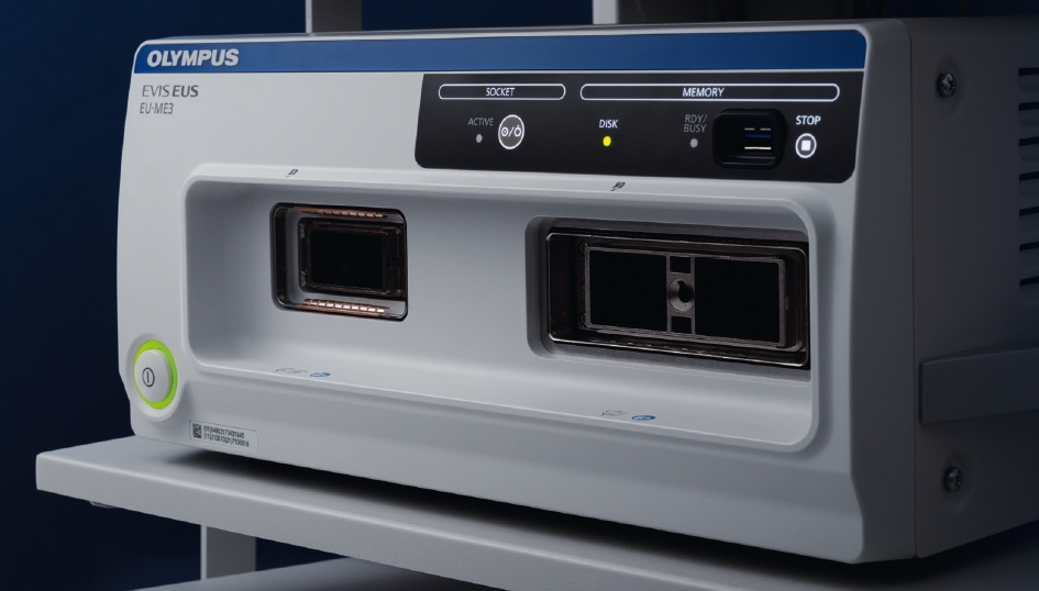
With more functions, better visualization, and enhanced operability, the EU-ME3 expands the dimensions of endosonography.
Brochure-
Enhanced B-mode
The EU-ME3 provides outstanding image quality and functionality – compatible to a high-end ultrasound center – in a compact body. B-mode image quality has been substantially
enhanced compared to our conventional processor (EU-ME2).Tissue Harmonic Echo (THE) mode
When ultrasound waves are propagated through tissue, distortion is produced and harmonic components are generated. The Tissue Harmonic Echo (THE) mode uses these components to build an image of the targeted area, providing a more detailed granular depiction. Advantages of harmonic imaging include improved resolution,improved signal-to-noise ratio, and fewer artifacts.
Improved Elastography
The EU-ME3 features an elastography function which visualizes the amount of strain in the tissue (tissue stiffness) during compression and retraction, making it possible to obtain more information about tissue properties.
Doppler Modes
The EU-ME3 offers three basic Doppler modes to distinguish blood flow more clearly - Color Flow, Power Flow, and Pulsed Wave Doppler (PWD). Doppler modes can be used to support safer procedures, benefitting both the patient and the physician.
In addition to the three basic Doppler modes, the EU-ME3 also features H-Flow. H-Flow is a more sensitive Doppler mode that shows directional blood flow with less blooming. It is especially useful for imaging small vessels around the tip of the echoendoscope.Contrast Harmonic Echo (CHE)
Contrast Harmonic Echo (CHE) images harmonic components from ultrasound contrast agents.
The newly added C-THE mode images signals from biological tissue and the contrast. -
Shear Wave Quantification (SWQ)
SWQ provides an absolute value of tissue stiffness within a region of interest. It performs this quantitative tissue assessment by calculating the propagation velocity of shear waves, generated from a push-pulse.
Elastography (i-ELST)
i-ELST is a new technology incorporated into the EU-ME3 that makes it easier to display elastic images, even when displacement due to pulsation is modest.
s-FOCUS
The EU-ME3 is equipped with an s-FOCUS mode that reduces the changein resolution with distance from the ultrasound transducer surface. s-FOCUS eliminates the need to manually adjust the focal zones during the procedure.
Wide Range of Compatibility
Integrating both electronic and mechanical scanning technologies, the EU-ME3 is compatible with echoendoscopes and miniature probes, creating a total endosonography solution for a full
range of applications.Customizable Features
Software options are available to meet the needs of any facility. Because the functions are optional, you can select and add the necessary functions according to your needs
and budget. -
Power Supply Voltage 220 – 240 V AC Voltage fluctuation Within ± 10% Frequency 50/60 Hz Frequency fluctuation Within ± 1 Hz Consumption electric power 370 VA Size Dimensions Main unit 371 (W) x 175 (H) x 480 (D) mm
445 (W) x 184 (H) x 530 (D) mm (maximum)Ketboard 392 (W) x 39 (H) x 210 (D) mm Weight Main unit 21.5 kg Ketboard 2.5 kg Classification Type of protection against electric shock Class I Degree of protection against electric shock of applied part TYPE BF applied part where no classification mark appears, the device is a TYPE BF applied part. Degree of protection against explosion The Ultrasound Center should be kept away from flammable gases. TYPE BF Applied Part This instrument can safely be applied to any part of the body except the heart EMC This instrument complies with the standards listed as follows :
IEC 60601-1-2 : 2001
IEC 60601-2-37 : 2007
CISPR 11 of emission : Group 1, Class BUltrasound scanning format Mechanical scanning, Electronic scanning Mechanical Scanning Display mode B-mode Scanning Radial scanning Compatible equipment Mechanical radial scanning ultrasound endscopes, Miniature probe Usable frequencies C5, C7.5, C12, C20, 7.5, 12, 20 MHz Display range 2, 3, 4, 6, 9, 12 cm Image adjustment Gain, Contrast, STC, Enhance Display processing Rotation Rotatable Display area Full circle, bottom sector, top sector, scroll Direction Normal/Inverse Cine memory Maximum 160 frames, Cine review function 3D 3D display, MPR display Measurement Distance, Area, Circumstance Electronic Scanning Display mode B-mode, FLOW mode, PW mode, THE mode, CH-EUS mode, ELST mode Scanning Radial scanning, Curved linear array scanning Compatible equipment Electronic radial scanning ultrasound endoscope
Electronic curved linear array scanning ultrasound endoscopeUsable frequencies 5, 6, 7.5, 10, 12 MHz Display range 2, 3, 4, 5, 6, 7, 8, 9, 12 cm Image adjustment Gain, Contrast, STC, Enhance, Compound Display processing Display area Radial: Full circle, bottom sector, top sector, scroll
Curved linear array : ConvexDirection Normal/Inverse Display pattern Single-screen/Dual-screen Cine memory Over 600 frames storable depending on the conditions
Cine review functionFocus Auto Preset Near/Far Focus setting Focus locatiion adjustable, Focus number adjustable FLOW mode COLOR FLOW mode, POWER FLOW mode, H-FLOW mode PW mode B+PW, COLOR+PW, POWER+PW, H-FLOW+PW Measurement Distance, Area, Circumstance, PW measurement THE (Tissue Harmonic Echo) mode *1, *2 THE-P, THE-R CH-EUS
(Contrast Harmonic EUS) mode *1, *2Display pattern CH-B, CH-Color Preset (CH agent type) 2 types, adjustable (middle or low) Frequency selection 2 types, adjustable (CH-R or CH-P) TIC analysis Displays the change over time of the average brightness of each ROI ELST (Elastography) mode *2 Pressurization state guide Strain graph, Pressurization bar Strain ratio *3 Displays the amounts of the strain and their ratio in two areas Recording Data Data format Still image Bmp, Jpeg, 3dv Movie data *1, *2 Avi Ancillary Equipment Keyboard Keyboard with built-in trackball, LCD touch panel and LED backlit keys Recording device Video printer (color/monochrome), DVR Video system center Monitor display selection Endoscopic/Ultrasound image Picture-in-picture Displays the endoscopic image as PinP sub-display on the ultrasound image Patient data Shares patient data with the video system center *1 Only available on EU-ME2 PREMIER *2 Only available on EU-ME2 PREMIER PLUS *3 Not available in some areas
-
-

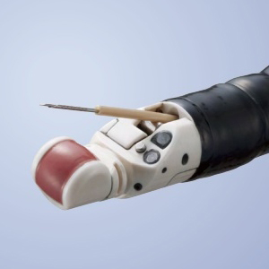
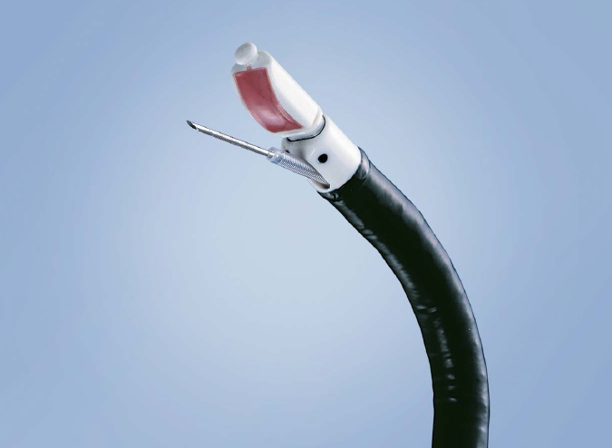
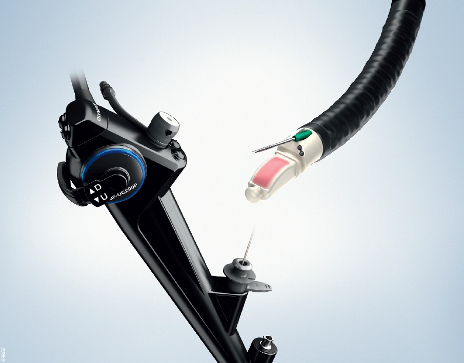
Linear Scanning Ultrasound EndoscopeGF-UCT260
Outstanding compatibility
Brochure
Excellent device handling -
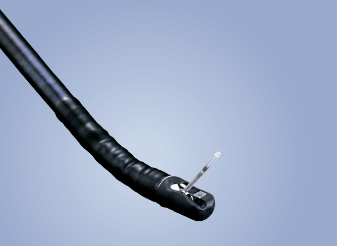
Radial Scanning Ultrasound EndoscopeGF-UE260-AL5
Maximises all-round examination
Brochure
Ideal for deep tissue examination
Wide angulation range -

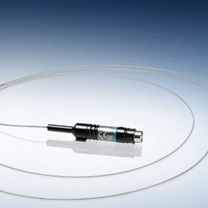
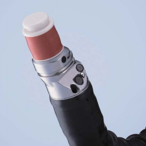
Miniature ProbeUM-S20-17S
Ultrasonic imaging via a slim endoscope
Brochure
Extra-slim probe design
360° ultrasonic view -
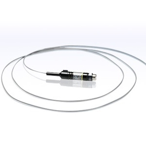
Miniature ProbeUM-3R
Ultrasound imaging via a standard endoscope
Brochure
Excellent 'all-rounder' ultrasonic probes
360° ultrasound view
-