RELATED
-
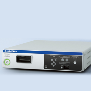
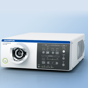
OTV-S190
-
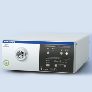
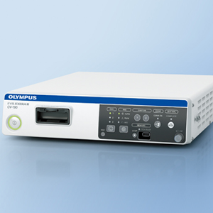
CLV-S190
-
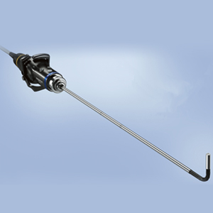
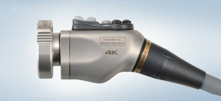
LTF-S190-5
-
-
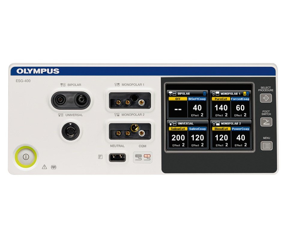
ESG-400
-
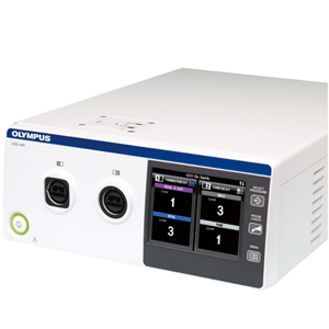
USG-400
-
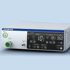
UHI-4
-
ENDOEYE HD II 10mm
-
ENDOEYE HD II 5mm
-

Laparoscopes HD 10mm
-
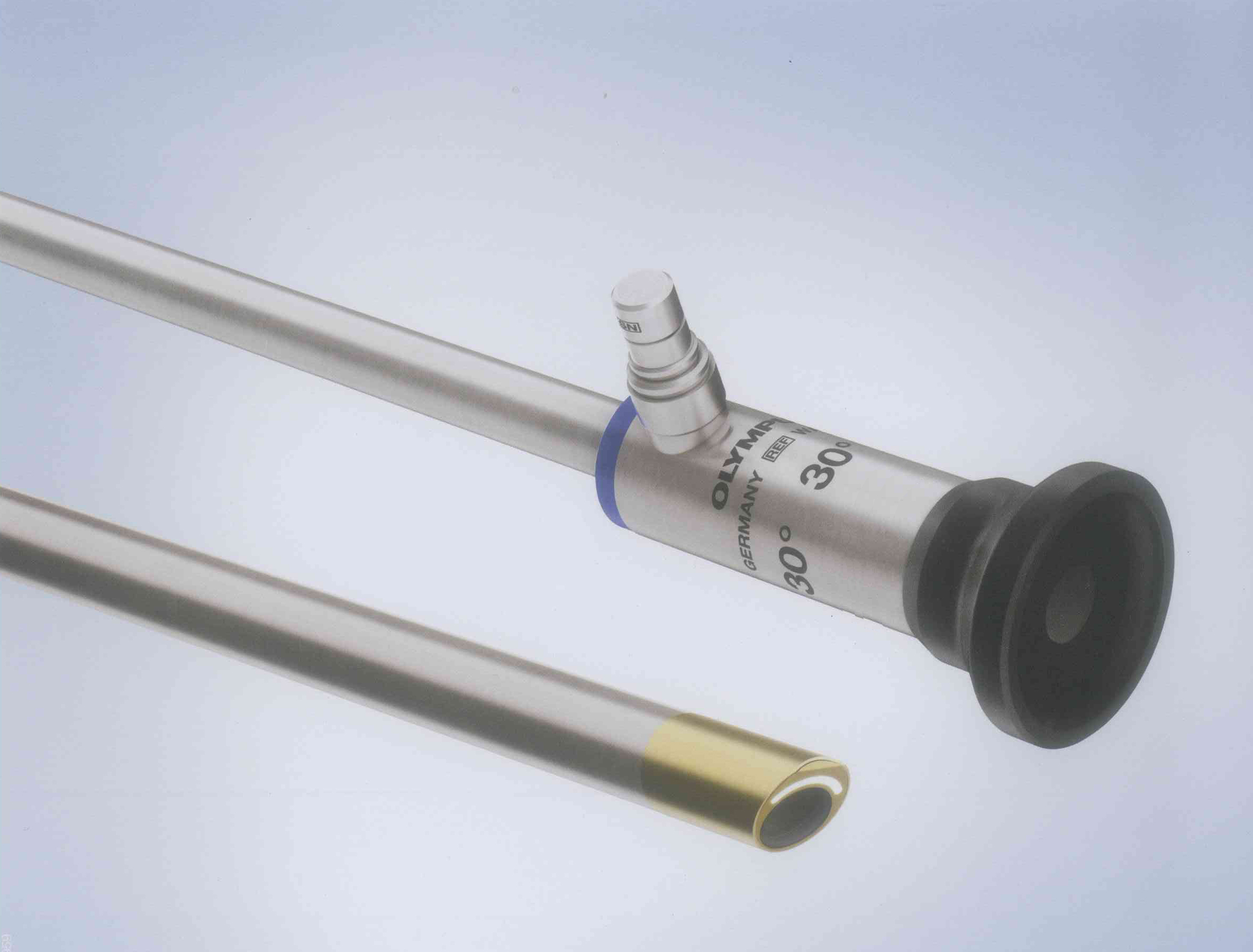
Laparoscopes HD 5mm
-

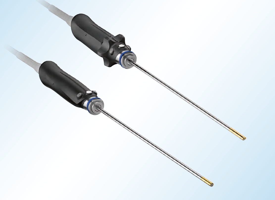
HiQ+ Monopolar Hand Instruments
-

HiQ+ Monopolar HF Electrode
-

HiQ+ Bipolar Hand Instruments
-

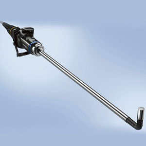
HiQ+ Suction and Irrigation Instrument
-
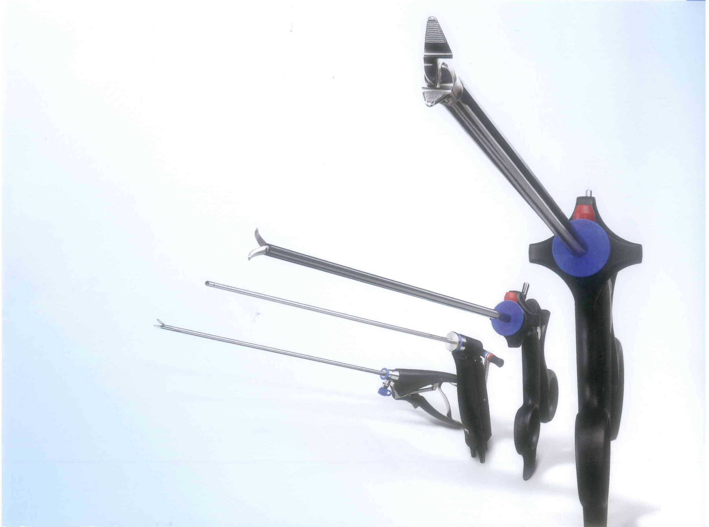
HiQ+ Needle Holder
-
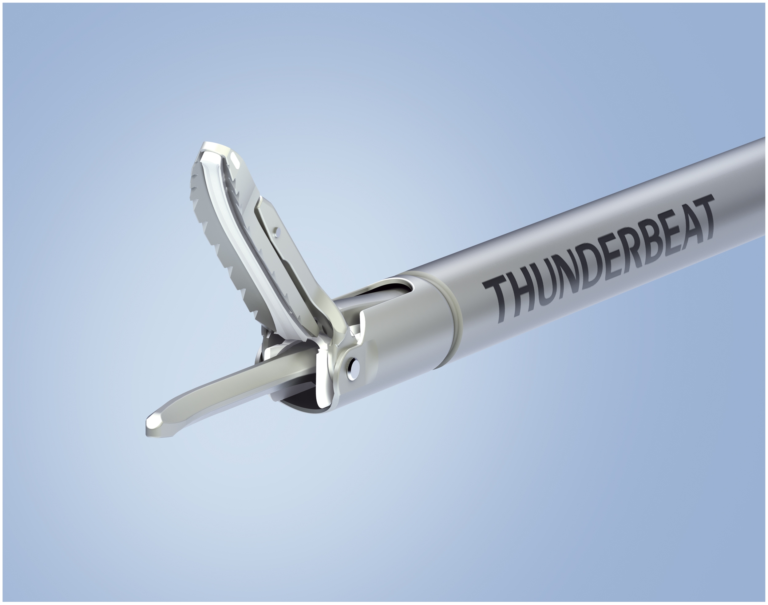
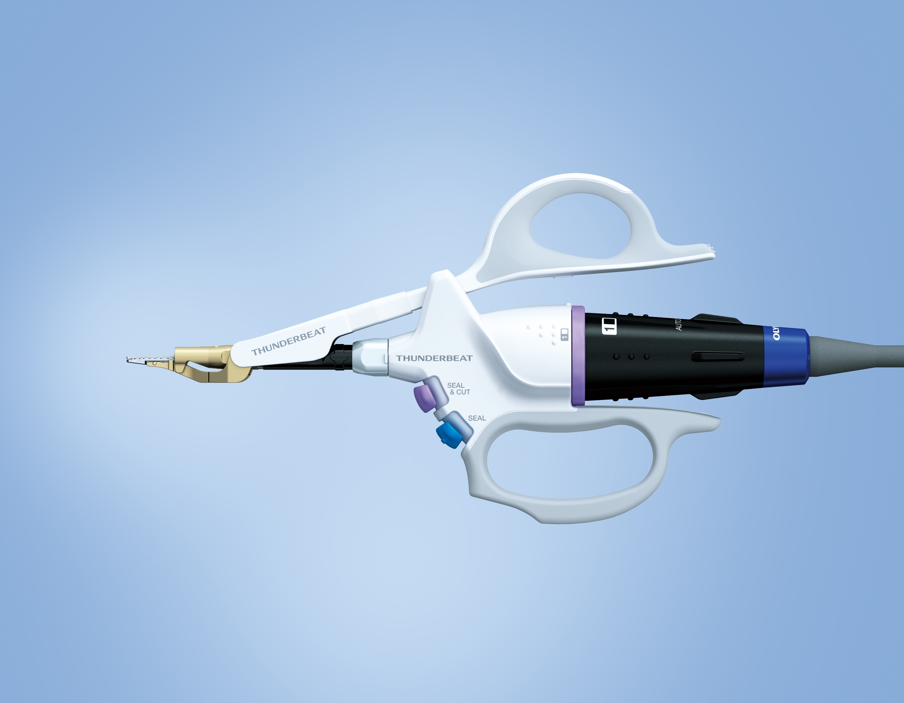
THUNDERBEAT
-
CLL-V1
-
OTV-S200
-
WA50080A/WA50082A
-
OTV-S700
-

CLL-S700
-
LTF-S190-10
-

POWERSEAL
-
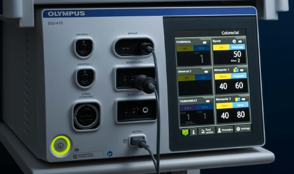
ESG 410
-
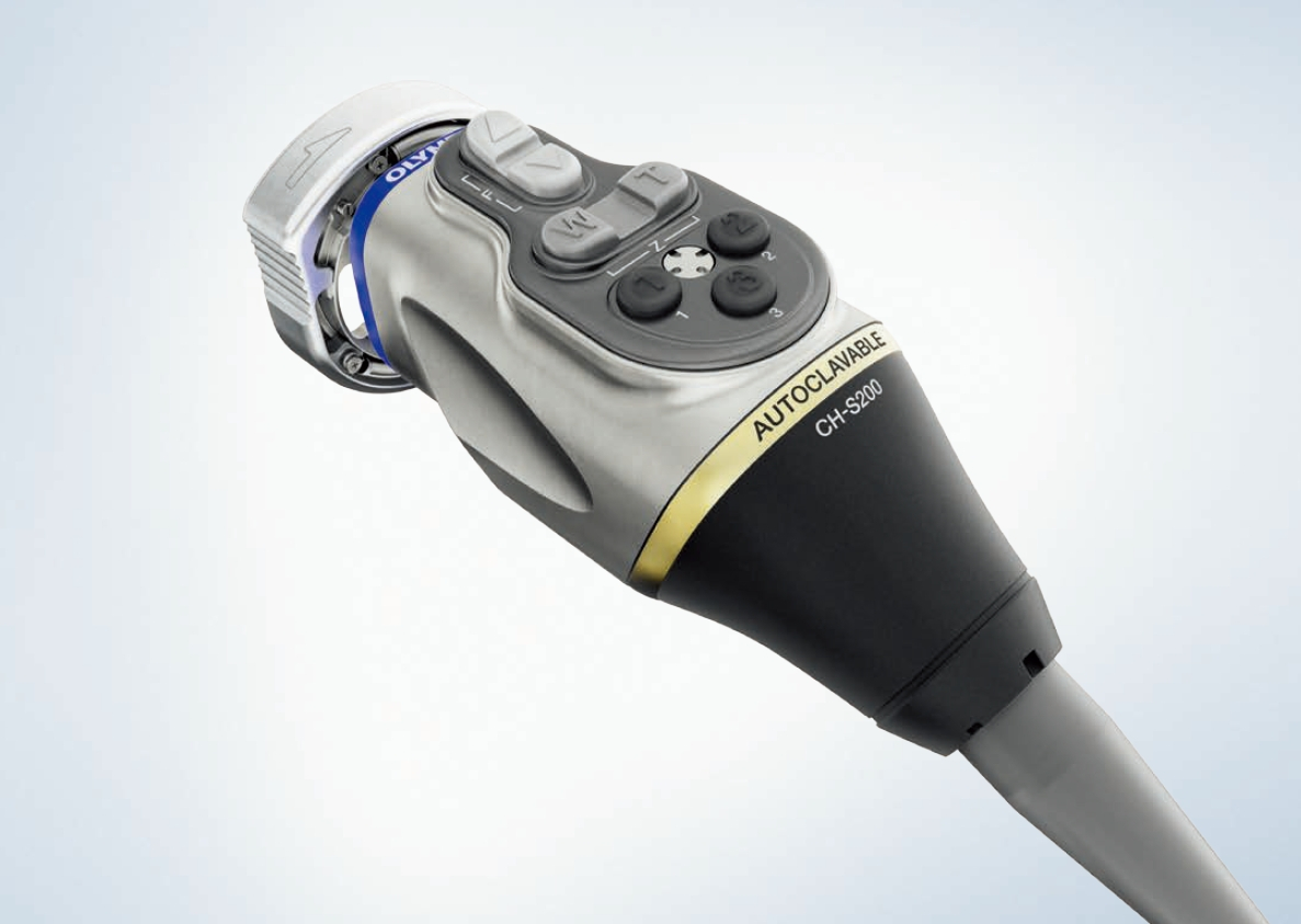
CH-S200-XZ-EA
-
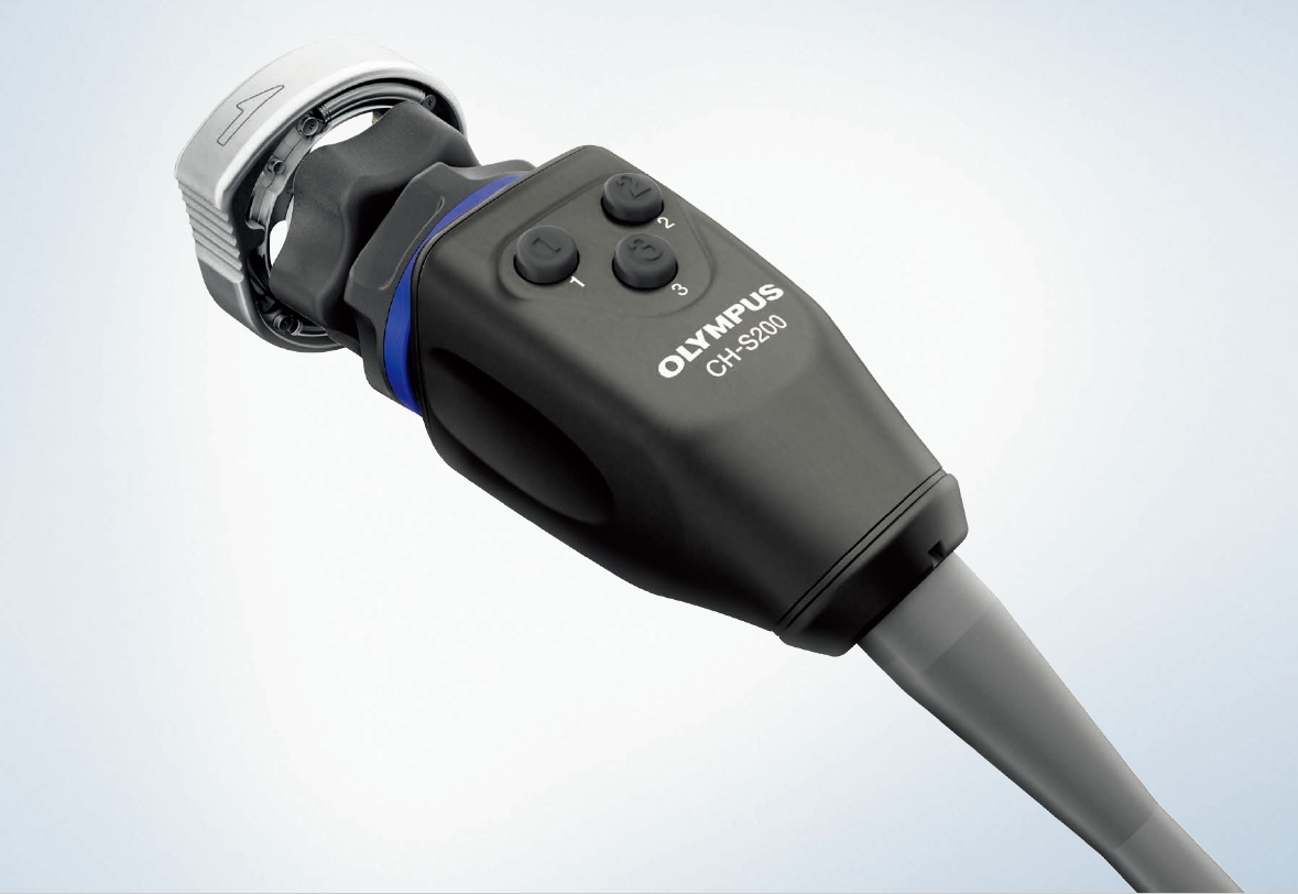
CH-S200-XZ-EB
-

CH-S200-08-LB
-
CH-S700-XZ-EA 4K ICG
-
CLV-190
-
CV-190